
Cryogenium
The next generation Cryo Plunger
Imaging of biological samples embedded in vitrified ice has become of great interest in recent years as it provides several advantages: the biological sample is in a fully hydrated state with superior preservation down to ultra-structural level, a vitrified sample is naturally compatible with the vacuum required for EM / Single Particle (SPT) / CLEM (Correlative Light and Electron Microscopy) and cryo-fluorescence provides very low photo-bleaching and high signal to noise imaging.
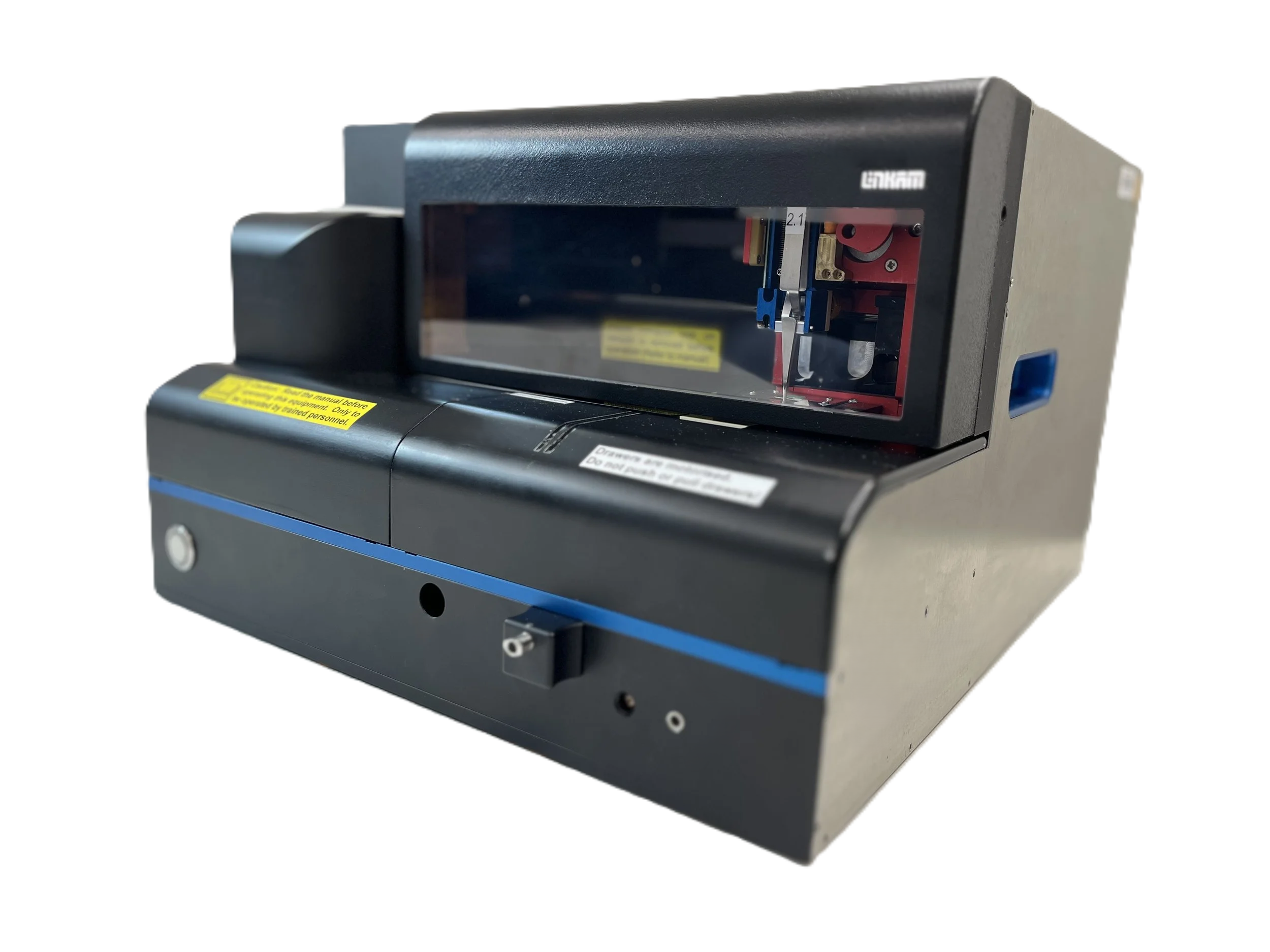
Preparation and handling of vitrified samples normally requires special skills and techniques. The novel design of the Linkam CryoGenium makes this a simple and reproducible process.
There are several common issues in a conventional plunge-freezing setup. Typically the sample is prepared on an electron microscopy grid and excess liquid is removed inside a humidity chamber using blotting paper to control the ice thickness of the frozen sample. The blotting paper usually needs alignment and blotting force calibration, while the paper does not work consistently over time because it soaks up moisture in the humidity chamber and prevents real-time optical monitoring during blotting.

The novel design replaces the common blotting mechanism to improve process stability. The system introduces real-time optical observation of the sample with control of the liquid film thickness. Film thickness is controlled by adjusting the speed at which the sample is drawn from the fluid as well as thinning of the film thickness by controlled suction. When the correct conditions are confirmed by the optical system, the user triggers plunging into a temperature-controlled bath of liquid ethane. Plunging is followed by automated loading into either a special cryo-holder or a cryo-transfer container. Fully automated, programmed operation is also possible.
The CryoGenium provides a platform that increases the success rate and quality of plunged grids, enabling improved efficiency and less time wasted at the TEM, resulting in cost savings.
Features / Highlights of CryoGenium
Fully Automated System
Automated grid preparation, sample loading, sample grid handling, and cryo-transfer in one compact unitReal-Time Optical Monitoring
Thin film interference enables water thickness to be monitored prior to plungingNovel system for control of film thickness
Water film thickness is accurately controlled by precisely controlled suction processIntegrated Glow Discharge
Fully controllable glow discharge unit integrated into the system, with oil-free vacuum pumpTemperature Controlled Ethane Bath
Temperature control of the Ethane bath ensures there is no solidification of the EthaneControlled Dew-Point of sample substrate
Dew-point control of sample support substrate inside the humidity chamber allows good stability and control of very thin and delicate liquid films on the sample supportControlled Temperature sample chamber
The temperature of the sample chamber can be controlled from 4°C to 40°C to ensure optimum conditions for the sampleAutoloading
Automated loading of the grid into cryo-transfer shuttle or cryo-storage box

The CryoGenium also features streamlined and easy-to-use software where the workflow, settings and operation of the plunger can be controlled from one place.
More information about the system can be found in the following Nature Communications paper.
https://doi.org/10.1038/s41467-022-30562-7
NEED MORE INFORMATION?
Contact us and one of our technical experts will be in touch shortly.
CryoGenium - The next generation of Cryo Plunger
CryoGenium integrates into your workflow
Single Particle Tomography (SPT)
SPT is growing rapidly and allows the determination of the molecular structure of proteins using EM and computational classification and reconstruction.
However, the sample quality and in particular the ice thickness have major influence on the signal-to-noise of the data. This is why the CryoGenium can become a key tool for SPT because it gives the user better control of ice thickness and sample preparation.
Cells, vesicles, and more
There is support for cartridges in the CryoGenium where sample grids can be seeded with cells, vesicles etc. and placed in an incubator. The whole cartridge is then placed into CryoGenium and the grids are picked up automatically and processed.
FIB-SEM and lamella preparation for cryo-EM tomography
Cells can be prepared, plunged, loaded into compatible cartridges for fluorescence or light microscopy screening in the Linkam CMS196 cryo-stage and then transferred to FIB/SEM to prepare lamellas.
The CryoGenium supports the CMS196 EM grid cassette, providing the ability to automatically place the plunged grid into the cassette and enabling a seamless workflow from bare grid to vitrified grid ready to be placed on the CMS196 for Cryo-CLEM work, or directly into the Cryo EM.
Why Cryo-CLEM?
Electron microscopy (EM) provides structural information at very high resolution. However, it gives only restricted insight into biological and chemical processes due to limitations in staining and sample preparation processes. Fluorescence microscopy on the other hand is a very sensitive method to detect biological, chemical and genetic processes and events inside living cells.
Cryo-CLEM brings it all together: it is a new and emerging technique that combines the individual advantages from both Fluorescence and EM by imaging the same sample location with both techniques and superimposing the complementary information.