Linkam Launches Enhancements to HFS Probe Stage

Expert in sample characterisation, Linkam Scientific Instruments, has unveiled its enhanced HFS600E-PB4 probe stage for use in micro-sized electrical component temperature control applications.


The new design was implemented in partnership with Linkam’s customer, Radiant Technologies, a major player in the design and manufacture of testing equipment for electrical systems. The combination of Linkam’s HFS600E-PB4 stage with Radiant test platforms has benefited Radiant’s customers for a number of years, allowing them to carry out analysis of macro-scaled electronic components including ferroelectric and piezoelectric materials across a temperature range from < -195 to 600 °C.

Continue Reading:

Linkam Paper at Microscopy and Microanalysis (M&M), Minneapolis, USA, July 2023

Linkam are pleased to announce that we will be presenting the following paper at the Microscopy and Microanalysis 2023 conference to be held in Minneapolis, USA July 23-27. Our poster will be on display on Monday, July 24, 2023, from 3:00 – 5:00 PM.

Plunger-robot for blot-free preparation of cryo-EM samples with controllable film thickness and real-time optical process control

Roman I. Koning¹, Hildo Vader², Martijn van Nugteren², Peter A. Grocutt², Wen Yang¹, Ludovic L. R. Renault¹, Abraham J. Koster¹, Arnold C. F. Kamp², Ivy Wang³, Maria Hariolaki³, Andrew Davies² & Michael Schwertner²

¹Electron Microscopy, Cell and Chemical Biology, Leiden University Medical Center, P.O. Box 9600, 2300 RC Leiden, The Netherlands
²Linkam Scientific Instruments Ltd., Unit 9, Perrywood Business Park, Honeycrock Lane, Salfords, RH1 5DZ, UK
*michaelschwertner@linkam.co.uk
³Diamond Light Source (B24) and Oxford University, UK

Figure 1: Comparison between the widely used workflow utilising pipetting and conventional paper-blotting (top row) and our proposed approach (bottom row) using sample-dipping and programmed suction with optical real-time monitoring.

Cryo-EM, cryo-tomography, Single Particle Tomography (SPT) and cryo-CLEM (Correlative Light and Electron Microscopy) have become standard tools for the investigation of protein 3D structures, cellular structures and more in the last decade. Despite the progress with cryo-EM instruments and automated sample loading and imaging the routine preparation of cryo-samples can be challenging.

Cryo-plunging techniques most widely used for vitrification are based on paper-blotting to adjust the thickness of the vitrified layer. We have identified this as a key drawback in the process because the blotting paper soaks up moisture while inside the high-humidity chamber leading to characteristics that are not always repeatable. As a solution we propose a novel method where fluid is removed for the sample thickness adjustment by means of a calibrated and programmed suction process. A comparison between conventional paper-based blotting and our proposed method is shown in Figure 1 and described in Ref [1] in more detail. Key benefits of the programmed suction are repeatability, straightforward automation and direct optical access to the sample for real-time monitoring and control of sample film thickness. The thin-film interference and the liquid surface meniscus characteristics observed in the real-time optical microscope allow to determine the best moment to plunge and represent a feedback loop to optimise film thickness.

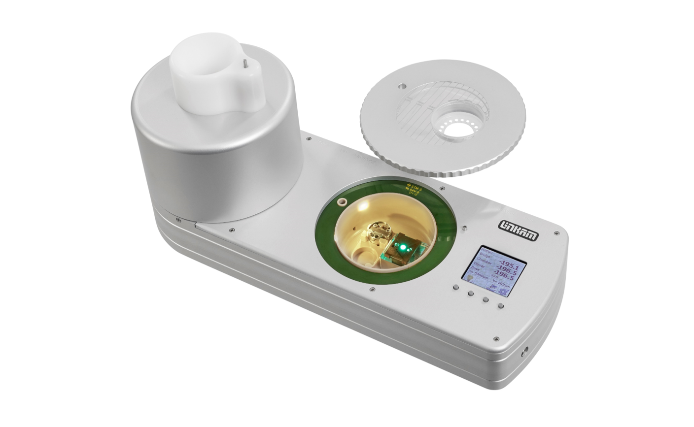
The overall setup of the plunger robot in the latest version is illustrated in Figure 2 and the principle is also detailed in [1]. The compact table-top unit contains an automated robotic tweezer arm, a glow-discharge unit to make the sample support hydrophilic, a grid box for sample support pickup, a humidity section with dew-point control, built-in microscope and suction unit for film thickness adjustment, a cup for liquifying and plunging into ethane and a location to deposit and cool the vitrified sample support. The device is computer-controlled via USB and automated sample preparation recipes can be run and configured.

Most importantly, the microscope image acquired immediately before plunging allows good prediction of the quality of the vitrified grid enabling pre-selecting or discarding of samples to make best use of EM beam time.

The setup improves repeatability due to a new principle, removes potential risks from manual sample handling and increases speed and throughput via automation. It can be used for both single-particle workflows as well as cell-based workflows.

Figure 2a: Automated robotic plunger system: Overview of main components with cryo-section on the left and humidity-section on the right.

Figure 2b: Detailed view of robotic plunger system with both drawer sections open with tweezer unit, suction system, ethane cup and glow-discharge unit visible.

References

[1] "Automated vitrification of cryo-EM samples with controllable sample thickness using suction and real-time optical inspection", R.I. Koning, H. Vader, M. van Nugteren, P. A. Grocutt, Wen Yang, L. L. R. Renault, A. J. Koster, A. C. F. Kamp & M. Schwertner, NATURE COMMUNICATIONS | (2022) 13:2985 | DOI: 10.1038/s41467-022-30562-7

Putting Meat to the Test: Imaging and Mechanical Testing Used to Understand the Properties of Meat Alternatives and How They Mimic Our Typical Meat Sensory Experience

An increasing number of consumers incorporate plant-based products and meat alternatives into their diet. Researchers at Reading Scientific Services Ltd. (RSSL) have performed tensile testing on both chicken and plant-based chicken alternative samples. Using a Linkam MFS stage, the team has assessed the failure point and breaking pattern of each of the samples, to determine which plant-protein product behaved most similarly to the chicken sample. This research offers a path for manufacturers in the meat alternatives industry to better understand the textural differences between chicken and alternative products, and can help inform future new product development for manufacturers looking to expand their product offering.

Continue reading at:

Linkam present new cryo-electron microscopy research at MC 2023

Linkam Scientific will present a new cryo-electron microscopy (cryo-EM) research paper at the Microscopy Conference 2023 (MC2023), Darmstadt, Germany: ‘New principle and robot for vitrification of cryo-EM samples with controllable sample thickness and real-time optical inspection’.

Continue reading at:

Improving measurement of electronic materials to boost device performance

Photovoltaics, ferroelectrics, and piezoelectrics have a suite of remarkable properties, with a long history of electrical applications in everything from ultrasound transducers to solar cells. But there is constant pressure to improve performance, and in this article, we look at how precisely controlling the temperature at which electrical measurements are made is helping researchers to improve their understanding of these materials.

Continue reading at:

Investigating the properties of meat alternatives and the sensory experience

Currently around 14% of UK adults (7.2 million) are following a meat-free diet, with 8.8 million pledging to go meat-free in 2022. Combined with the increasing popularity of the flexitarian diet – a portmanteau of vegetarian and flexible, consumption of meat alternatives is rising. The pressure for meat alternatives to fulfil the expectations of former meat eaters has increased.

Read More:

Temperature-controlled measurements are helping scientists make better electrical insulators

Perovskites are a class of materials with great promise in electronics, thanks to their wide array of tuneable dielectric* (insulator)  properties. However, these properties hinge on the presence or absence of inherent structural defects and impurities, as well as on environmental conditions such as temperature. Understanding these influences is critical for developing better-performing electronic components. Here is a summary of how temperature-controlled analysis can be used to bring out these properties.

Continue reading at:

Cryomicroscopy: The coolest technique in pharma research

In the last two decades, cryomicroscopy methods have become widespread and effective tools used by life scientists, pharmaceutical researchers, and more, to examine biological structures close to their native state.1 The capabilities of cryomicroscopy to visualize the structure of biological molecules, such as proteins and protein complexes, make a valuable addition to existing methods such as X-ray crystallography and nuclear magnetic resonance (NMR). Determining the structure of proteins and protein complexes, which are of particular interest as drug targets and are the subject of investigation to gain a deeper understanding of disease mechanisms, is an important element of drug discovery.

在过去的二十年中,冷冻显微镜方法已经成为生命科学家、制药研究人员等广泛使用的有效工具,用于检查接近其原生状态的生物结构1。冷冻显微镜能够可视化蛋白质和蛋白质复合物等物质的生物分子结构,是对现有的方法如x射线晶体学和核磁共振(NMR)等的有价值的补充。确定蛋白质和蛋白质复合物的结构是药物发现的一个重要部分,这对研究药物靶点非常有意义,也是深入了解疾病机制的重要课题,。

Read more (Chinese only):

Instrument.com.cn

bio-equip.com

Temperature-controlled measurements are helping scientists make better electrical insulators

Perovskites are a class of materials with great promise in electronics, thanks to their wide array of tuneable dielectric (insulator)  properties. However, these properties hinge on the presence or absence of inherent structural defects and impurities, as well as on environmental conditions such as temperature. Understanding these influences is critical for developing better-performing electronic components.

Read More:

Fierce Electronics

Linkam Scientific Instruments Announces New NEXUS Software

Linkam Scientific Instruments has launched new software, NEXUS, to control and record all aspects of heating, cooling, vacuum, humidity and other environmental conditions in a Linkam stage with optional optical measurement capabilities. NEXUS Core is the main NEXUS platform that introduces a range of new specifications to enable a greater degree of experiment control. Users can easily monitor the status of their experiment with the live display feature, quickly creating observations and adjustments that alter the data collection process. The upgraded software ensures precise synchronisation between samples through the live status display in parallel with a real-time temperature chart.

Read More:

Linkam Scientific Instruments mark 40th anniversary in new UK premises

Linkam Scientific Instruments has opened the doors at its new UK premises in Redhill, Surrey, UK, on the occasion of its 40th anniversary in temperature-controlled microscopy. The move stems from the company’s unprecedented growth in recent years, particularly in custom-designed solutions to support scientific research in precision temperature- and humidity-controlled environments. Now celebrating its 40th year, Linkam has gone from strength to strength, not only with the new site expansion, but also by growing its team with two new hires in its sales and marketing department and human resources.

Read More:

The Importance of Temperature Control in Pharmaceutical Analysis

In many areas of the pharmaceutical industry, it is becoming increasingly important to understand the micro- and thermo-mechanical properties of a substance. Temperature-controlled microscopy covers a wide range of analytical techniques, including methods that involve combining traditional thermo-analytical methods, such as differential scanning calorimetry (DSC), and image analysis techniques, such as thermal analysis by structural characterisation (TASC), to characterise pharmaceuticals and their component materials.

Read More:

New Development in Electron Microscopy Grid Vitrification

Linkam previewed its new cryo plunger—CryoGenium—an exciting new development in electron microscopy (EM) grid vitrification. Imaging of biological samples embedded in vitrified ice has become of great interest in recent years as it provides several advantages: the biological sample is in a fully hydrated state with superior preservation down to ultra structural level, a vitrified sample is naturally compatible with the vacuum required for EM/single particle (SPT)/correlative light and electron microscopy (CLEM), and cryo fluorescence provides very low photo-bleaching and high signal to noise imaging.

Read More: